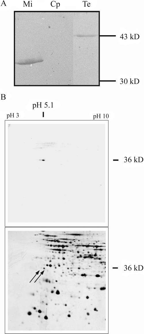
Figure 2.
Protein gel electrophoresis and subsequent western-blot analysis of total and organellar extracts. A, Mitochondria (Mi) were purified from Arabidopsis cell cultures, chloroplasts (Cp) were isolated from green Arabidopsis plants, and total soluble protein extracts (Te) were also obtained from green Arabidopsis plants. The proteins were separated by one-dimensional gel electrophoresis, blotted, and the membranes were incubated with an antibody directed against the AtStr1 protein. B, Mitochondria were purified as described above. Their proteome was separated by two-dimensional gel electrophoresis and analyzed by western-blot analysis as described above (top). The corresponding protein spots marked by arrows were localized on a Coomassie-stained gel that was run in parallel. The spots were cut and the proteins were analyzed by mass spectrometry.

