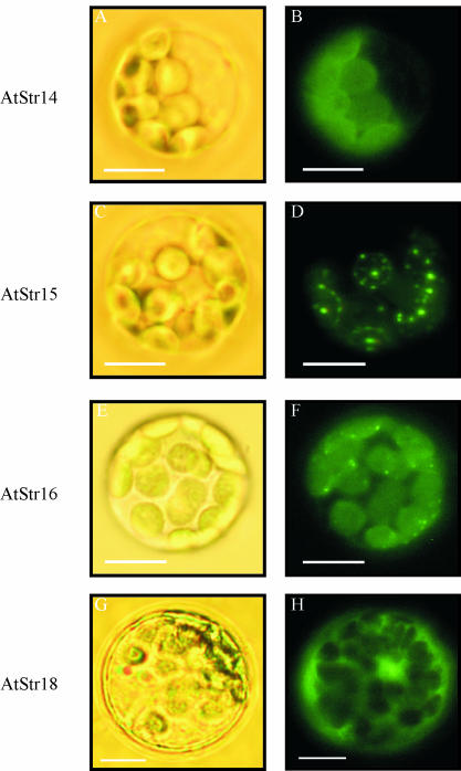
Figure 4.
Targeting analysis of all members of the AtStr group VI visualized by fluorescence microscopy. The fusion constructs of AtStr14, AtStr15, AtStr16, and AtStr18 with pGFP-N were introduced into Arabidopsis protoplasts. The protoplasts were incubated overnight at room temperature and then analyzed with an Axioskop microscope with filter sets optimal for GFP fluorescence (BP 450–490/LP 520). Bright field images (A, C, E, and G) were made to visualize the protoplast's cell membrane and chloroplasts. Fluorescence images of the same protoplasts are shown in B, D, F, and H.

