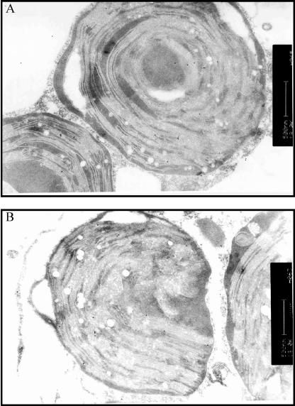
Figure 6.
Immunogold localization of GFP fusion protein in transiently transformed protoplasts by transmission electron microscopy. A, Chloroplast with numerous (>12) gold particles (i.e. GFP immunoreactivity) close to another chloroplast with and to a mitochondrion (below) without immunogold label. B, Chloroplast at the left with about 12 gold particles at the thylakoid membrane. The mitochondrion at the right has no label. The chloroplast at the right contains only one label at the thylakoid membrane. The plasma membrane of the protoplast has one gold label. The scale bars represent 500 nm.

