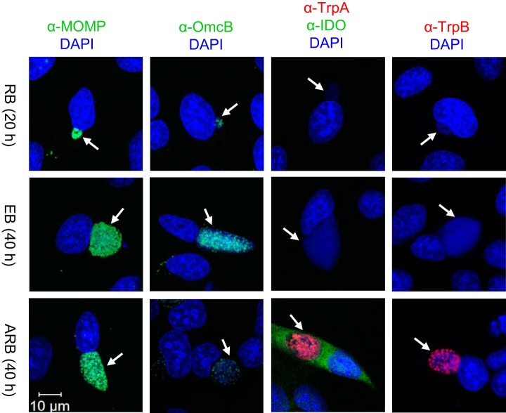
Fig. 1.
Confocal microscopy images of HeLa cells infected with C. trachomatis serovar D/UW-3/CX cultured in supplemented RPMI medium in the absence (RB, EB) or presence of IFN-γ (ARB). Either 20 or 40 h post infection cells were fixed and stained with antibodies for MOMP (green), OmcB (green), IDO (green), TrpA (red) or TrpB (red), as well as DAPI (blue) to detect host and bacterial DNA. White arrows indicate the position of inclusions and the white scale bar indicates 10 μm.

