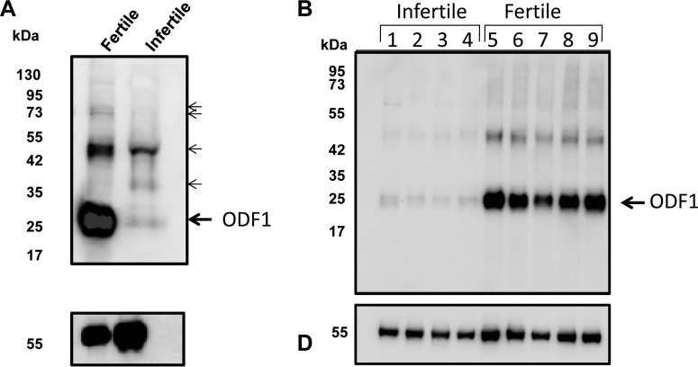
Fig. 2.
Severe reduction in ODF1 from the gametes of the infertile male. (A) Western blotting analysis. Proteins were extracted from either a known fertile donor (lane 1) or the infertile male (lane 2). The proteins were run into SDS-PAGE, then transferred to nitrocellulose. The membrane was then probed for using anti-ODF1 antibody (top panel). After this, the membrane was stripped and reprobed using anti β-tubulin antibody (lower panel). The position of the molecular weight markers are shown on the left-hand side. The large arrow shows the expected position of ODF1. The smaller arrows higher molecular mass cross-reacting bands that were excised for nano-LC-MS/MS analysis. (B) Immunoblot using the anti-ODF1 against suspected idiopathic infertile men (lanes 1–4) or control fertile donors.

