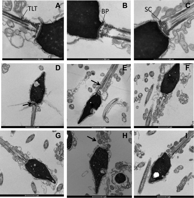
Fig. 5.
Ultrastructure changes to the implantation plate of spermatozoa with reduced ODF1. The appearance of a normal implantation plate with fertile samples is shown (A–C). The thin laminated fiber (A;TLT), basel plate (B, BP), and striated columns (C, SC) are labeled. Within the several reduced ODF1 infertile sperm (D–I) a lack of the basel plate formation was observed (D, arrow), lack of or poorly developed striated columns (D–I), and absent or smaller than usual thin laminated fibers (D, E, H).

