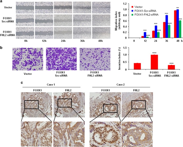
Figure 5.
Co-expression of FOXK1 and FHL2 is associated with metastatic phenotypes in human CRC. (a) For the wound-healing experiments, the cells were analysed with live-cell microscopy. Original magnification, × 10. ***P<0.01, ****P<0.001. (b) Vector, stable FOXK1 transfectants were transfected with FHL2 siRNA 48 h later, and the invasive ability of the cells decreased; ****P<0.001. The experiments were repeated at least three times. (c) Representative IHC images are shown for FHL2 and FOXK1 expression in lymph node metastatic cancer tissues. Scale bars, 100 μm in c. These pictures were representatives of three independent experiments with identical results.

