Figure 6.
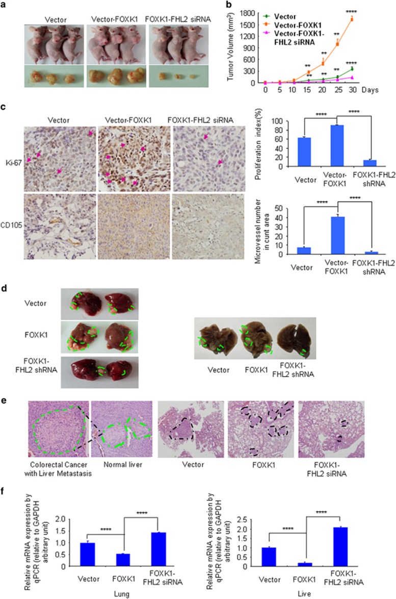
FOXK1 synergizes with FHL2 to promote tumour proliferation and metastasis in vivo. (a) Evaluation of tumorigenesis in nude mice subcutaneously injected with SW480-Vector, SW480-FOXK1 and SW480-FOXK1–FHL2-shRNA cells. Images were captured on day 30 after injection. (b) Tumour size was measured five days after tumour cell inoculation in each group. **P<0.05, ****P<0.001, vector vs FOXK1 and FOXK1 vs FOXK1–FHL2-shRNA, respectively. (c) FHL2 knockdown significantly inhibited FOXK1-induced proliferation (Ki-67, ****P<0.001, vector vs FOXK1 and FOXK1 vs FOXK1–FHL2-shRNA, respectively), and a considerable decrease of tumour vessel density (CD105, ****P<0.001, vector vs FOXK1 and FOXK1 vs FOXK1–FHL2-shRNA) was observed by IHC. (d) Mice were orthotopically transplanted with indicated cells (n=3 in each group). Representative images of metastatic loci in lungs or liver of blue dotted lines were shown. (e) The mice were killed, and metastatic cancer tissues were stained with haematoxylin and eosin (H&E). (f) The expression of E-cadherin in tumours derived from SW480 cells was determined by qPCR. ****P<0.001, vector vs FOXK1; FOXK1 vs FOXK2-FHL2-siRNA. Scale bars represent 100 μm in c and 200 μm in d.

