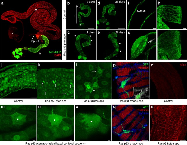
Figure 2. Targeting quadruple combinations to the adult hindgut.
(a) The adult Drosophila digestive track. Hindgut cells are visualized with byn>GFP; nuclei are in red. (b–i) Control (byn>GFP,dcr2) and rasG12V p53Ri ptenRi apcRi hindguts 7 and 21 days after induction. Asterisks in c and e indicate regions of multilayering. Longitudinal optical sections (f,g) and pylorus regions (h,i) are shown. (j–o) Control (j) and rasG12V p53RiptenRi apcRi (k–o) ilea; arrows indicate migrating cells. (l) Close-up view view of k. (m–o) Apical-to-basal confocal sections of a migrating cell (asterisk). (p,q) Surface views of rasG12V p53Ri smad4Ri apcRi hindguts with cells migrating on top of the muscle layer. (p) Inset: laminin (grey) and GFP channels only to highlight trachea (arrows). (r,s) Phospho-Src staining of control and rasG12V p53Ri ptenRi apcRi hindguts.cr, crop; h, hindgut; i, ileum; m, midgut; mp, malphigian tubules; p, pylorus; r, rectum; t, trachea. Scale bars, 250 mm (a–e) and 25 mm (f–s).

