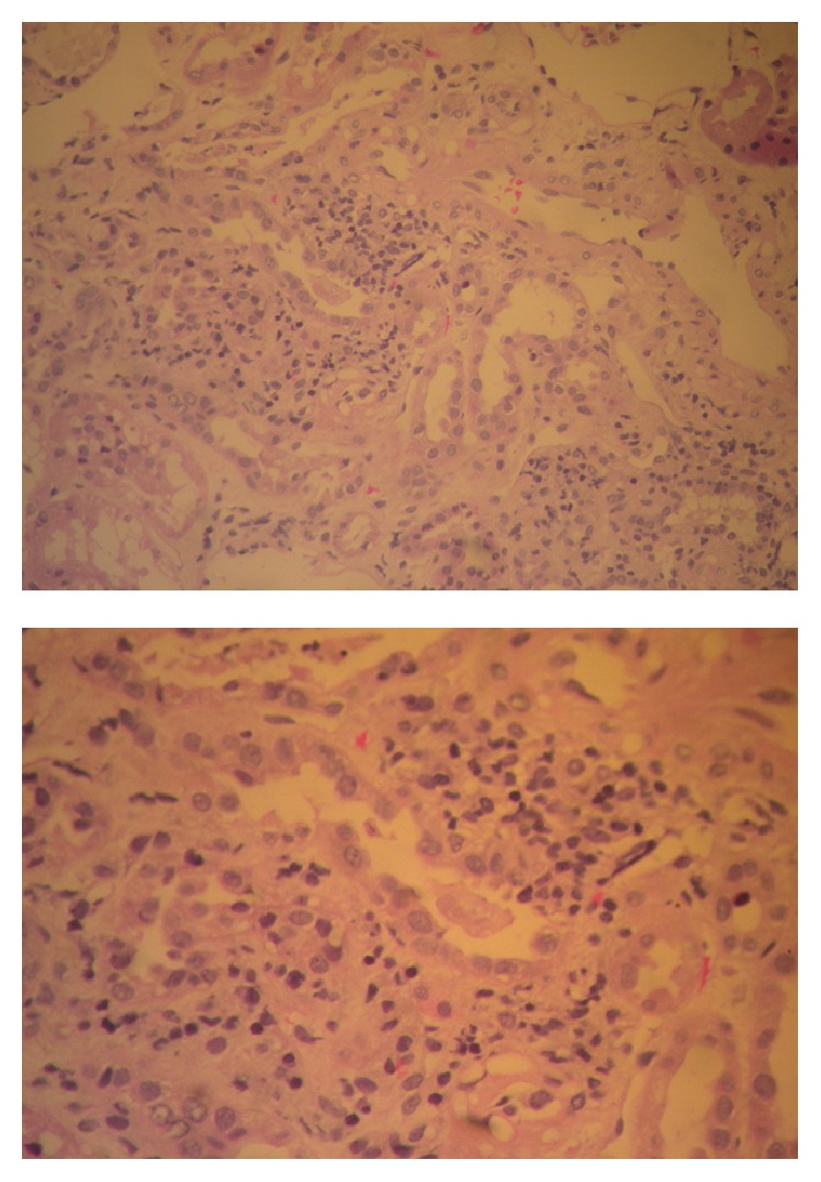
Figure 2.

Renal biopsy: findings from light microscopy showing evidence of AIN with interstitial edema and inflammatory peritubular infiltrations composed mainly of lymphocytes. The glomeruli have evidence of nonspecific mild mesangial enlargement, with no abnormalities of the glomerular basement membrane.
