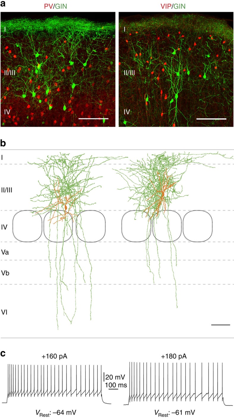
Figure 1. LII/LIII GIN cells show typical characteristics of Martinotti cells.
(a) Fluorescent staining of triple transgenic mice (left: PV-cre::tdTomato::GIN, right: VIP-cre::tdTomato::GIN) used for the present experiments. In all, 50-μm-thick frontal sections from the barrel cortex are shown. PV or VIP cells, respectively, are labelled red and GIN cells green. Layers are indicated as I–IV. Scale bar, 100 μm. (b) Neurolucida reconstructions of LII/LIII biocytin-filled GIN cells. Somatodendritic compartments are shown in orange and axonal arborizations in green. Note the dense axonal branching in LI, which is characteristic for MC. Layers are indicated as I–VI. Scale bar, 100 μm. (c) Whole-cell current-clamp recordings of GIN cells shown in b. Depolarizing current injections caused an adapting firing pattern in these cells, as it is typical for MC.

