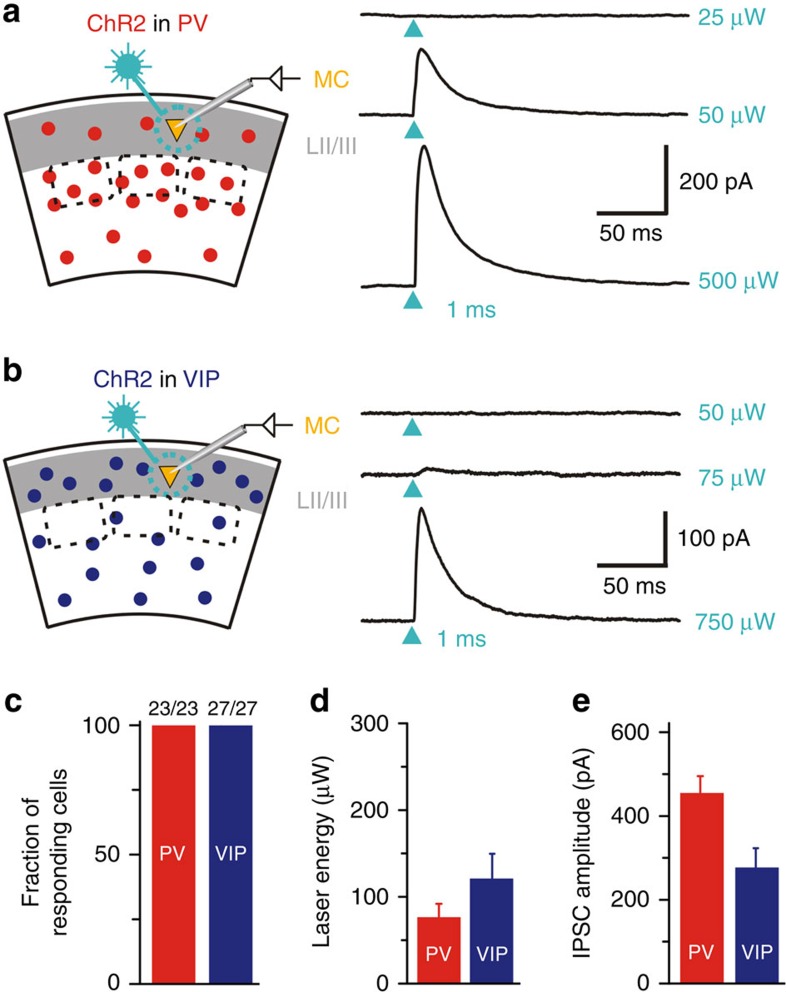
Figure 2. PV and VIP cells reliably target MC in LII/LIII of S1.
(a,b) Left: schematic of recording configuration for photostimulation of ChR2-expressing PV or VIP cells while recording from LII/LIII MC. Right: Examples of photostimulation-induced inhibitory postsynaptic currents (IPSC). Arrowheads indicate photostimulation (473 nm laser, 1 ms) of PV interneurons (a) or VIP interneurons (b) at three different intensities (subthreshold, threshold and 10 × threshold). (c) Proportion of MC responding to photostimulation of PV and VIP cells. In both experimental designs, the success rate was 100%. This indicates that each MC receives inhibitory input from PV and VIP cells. (d) Threshold laser energy to elicit IPSC in MC by photostimulation of PV and VIP cells. Note the same range (mean±s.e.m) for both groups. (e) Mean±s.e.m. of IPSC amplitudes (at 10 × threshold level) in MC for each group (PV: n=16, age P38–P60, VIP: n=18, age P35–53). Optical stimulation of the PV cell population tends to result in larger multicomponent IPSC amplitudes.

