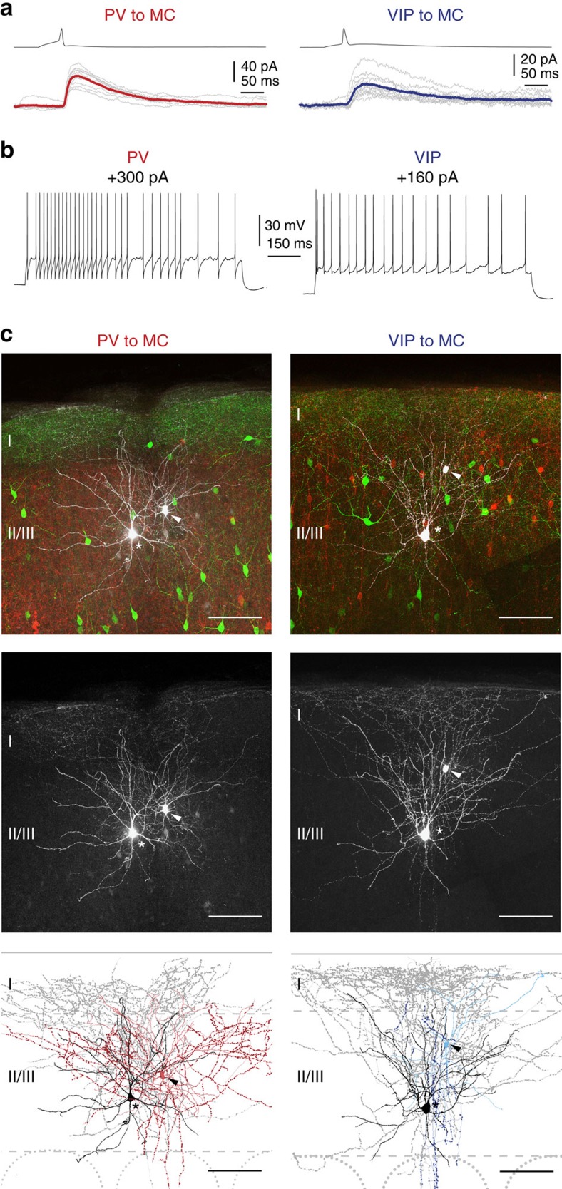
Figure 3. Electrophysiology and morphology of LII/LIII PV–MC and VIP–MC pairs.
(a) Examples of connected pairs of presynaptic PV or VIP cells and postsynaptic MC in LII/LIII. The average of 10 individual IPSCs (grey traces, evoked by repetitive stimulation) is shown in colour (PV to MC: red, age P23; VIP to MC: blue, age P27). Presynaptic spikes reliably evoke IPSCs in both cases. (b) Whole-cell recordings of a presynaptic PV (left) and a VIP cell (right). During depolarizing current injections, the PV cell shows a fast spiking pattern, whereas the VIP cell shows an adapting firing pattern. (c) Staining of acute brain slices containing morphologically recovered and synaptically connected pairs as well as the corresponding Neurolucida reconstructions (left: PV to MC, right: VIP to MC). The connected cells are shown in white (pseudo-coloured). Asterisks mark MC somata, arrowheads somata of presynaptic cells. GIN cells are labelled green and the corresponding presynaptic population (PV or VIP) is labelled red (tdTomato-fluorescence). For clarity, connected cells are shown separately as grey-scale images in the middle. The reconstructed pairs are shown at the bottom. Soma and dendrites of GIN cells are labelled black and the corresponding axon grey. The recorded PV cell exhibits a multipolar dendritic morphology (light red) and a locally dense axon (red), as described for basket cells. The VIP cell shows an atypical tripolar dendritic configuration (light blue) and an axon (blue) descending towards the white matter. Complete reconstructions are displayed in Supplementary Fig. 4. Scale bars, 100 μm.

