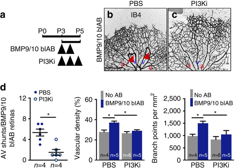
Figure 5. PI3K inhibition prevents AVM formation in BMP9/10 blAB-treated mice.
(a) Schematic representation of the experimental strategy to assess the effects of PI3K inhibition on the retinal vasculature of BMP9/10 blAB injected mice. Arrowheads indicate the time course of injection of BMP9/10 blAB and PI3Ki or PBS. (b,c) IB4 staining of retinal flat mounts from mice treated with BMP9/10 blAB (b) and BMP9/10 blAB and PI3Ki (c). Red arrowheads in b indicate AV shunts. Scale bars, 200 μm in b,c. (d) Quantification of AV shunt number, vascular density and number of branch points. n=4–6 Retinas per group. Each dot represents one retina. Error bars: s.e.m., *P<0.05, Mann–Whitney U-test. a, artery in red; v, vein in blue.

