Figure 3.
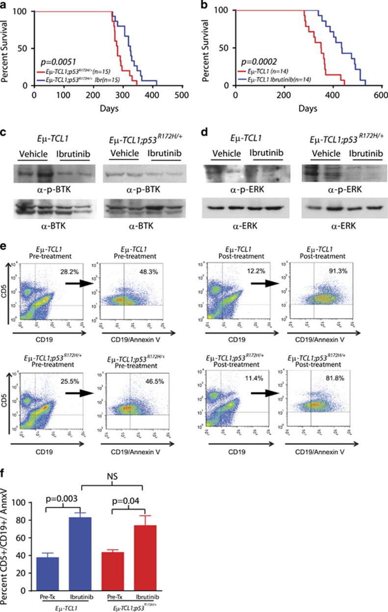
Ibrutinib prolongs survival and activates downstream pathways independent of p53. (a) Kaplan–Meier curves of vehicle- and ibrutinib-treated Eμ-TCL1;p53R172H/+ mice. (b) Kaplan–Meier curves of vehicle- and ibrutinib-treated Eμ-TCL1 mice. (c) Western blot of total BTK and phospho-BTK-(Tyr223) levels in spleens from short-term ibrutinib-treated Eμ-TCL1 and Eμ-TCL1;p53R172H/+ mice. (d) Western blot of total ERK and phospho-ERK (Thr202/Tyr204) levels in spleens from short-term ibrutinib-treated Eμ-TCL1 and Eμ-TCL1;p53R172H/+ mice. (e) Flow cytometry data demonstrating CD5+/CD19+/AnnexinV+ cells in the peripheral blood of Eμ-TCL1;p53R172H/+ and Eμ-TCL1 mice pre and post short-term ibrutinib treatment. Cells were initially gated by their expression of CD5, CD19 and CD5/CD19 and then further analyzed by their uptake of Annexin V. (f) Bar graph representing the percent of CD5+/CD19+/AnnexinV+ cells in the peripheral blood of Eμ-TCL1;p53R172H/+ (n=4) and Eμ-TCL1 (n=4) mice pre- and post-ibrutinib treatment.

