Figure 6.
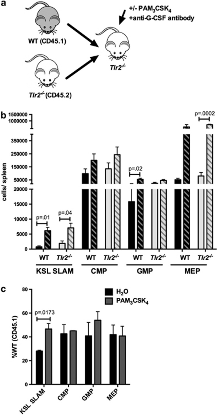
Cell-autonomous TLR2 signaling contributes to PAM3CSK4-mediated HSC expansion. (a) Chimeric animals were generated by transplanting equal numbers of WT (CD45.1) and Tlr2−/− (CD45.2) bone marrow cells into lethally irradiated Tlr2−/− (CD45.2) recipients. (b) After allowing time for engraftment (12 weeks), recipients were treated with PAM3CSK4 (100 μg intraperitoneally (i.p.) q48 h × 3 doses) and a G-CSF neutralizing antibody and the absolute numbers of KSL SLAM, common myeloid progenitor (CMP), granulocyte–monocyte progenitor (GMP) and megakaryocyte–erythroid progenitor (MEP) cells per spleen for each donor cell type were determined by flow cytometry. Shown are cells/spleen for the indicated populations. Dark bars indicate WT (CD45.1) cells, and light bars indicate Tlr2−/− (CD45.2) cells. Solid bars indicate water-treated controls, and hatched bars indicate PAM3CSK4-treated mice. (c) The relative frequencies of wild-type donor (CD45.1) HSPCs are shown for PAM3CSK4-treated or water control-treated chimeric mice. Data represent three chimeras from each treatment group from three separate experiments, and P-values were determined by two-tailed Student's t-test. Error bars represent mean±s.e.m.

