Figure 1.
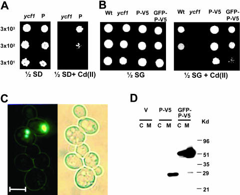
AtPcr1 overexpression confers Cd(II) resistance in yeast and AtPcr1 is localized in the plasma membrane. A and B, Growth in the presence or absence of Cd(II) of ycf1 cells carrying the empty pFL61 or pYES2/NTC vector (ycf1), the pFL61-AtPcr1 (P), the pYES2/NTC-AtPcr1-V5 (P-V5), or the pYES2/NTC-GFP-AtPcr1-V5 (GFP-P-V5) construct. The yeast strains were grown at 30°C for 4 d on 1/2 SD or 1/2 SG plates with or without 50 μm Cd(II). As a control, an isogenic wild-type (wt) strain was also examined. C, Localization of AtPcr1 in cells overexpressing GFP-AtPcr1-V5. Left, a fluorescent image of GFP-AtPcr1-V5-expressing yeast cells. Right, a bright field image of the same cells shown in the left panel. Bar represents 5 μm. D, Presence of AtPcr1 in the membrane fraction of AtPcr1-V5 (P-V5) and in both the membrane and cytosolic fractions of GFP-AtPcr1-V5 (GFP-P-V5)-expressing ycf1 cells grown on SG medium. The microsomal and cytosolic fractions were isolated and their proteins (50 μg each) were electrophoresed on SDS-PAGE and detected by the V5 tag antibody. The predicted AtPcr1-V5 and GFP-AtPcr1-V5 sizes are 25 and 50 kD, respectively.

