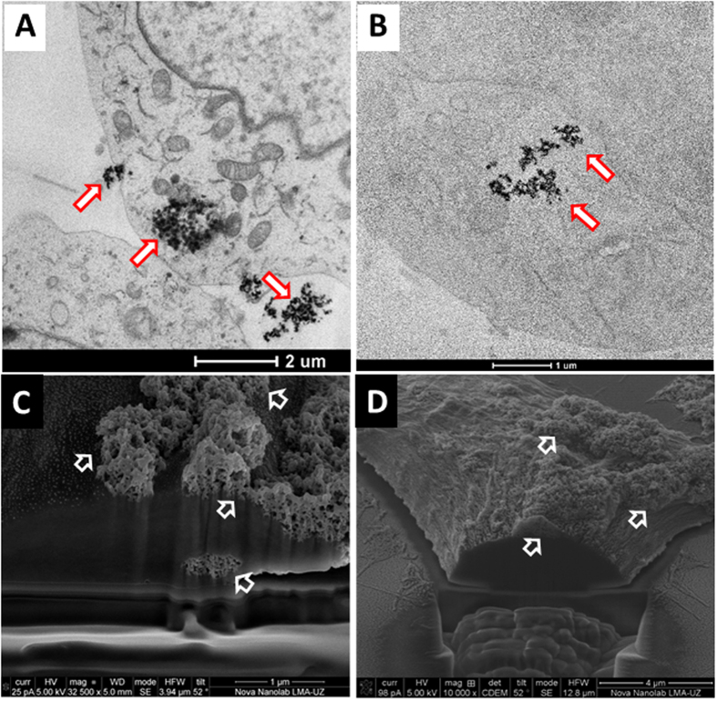
Figure 4.
TEM (A,B) and FIB-SEM (C,D) images of SH-SY5Y cells incubated with PEI-MNPs and PAA-MNPs for 24 hours. The TEM images of (A) PEI-MNPs and (B) PAA-MNPs show that the internalized particles formed clusters. The FIB-SEM images of the same cells show the presence of dense agglomerates of (C) PEI-MNPs and (D) PAA-MNPs attached to and crossing the cell membrane.

