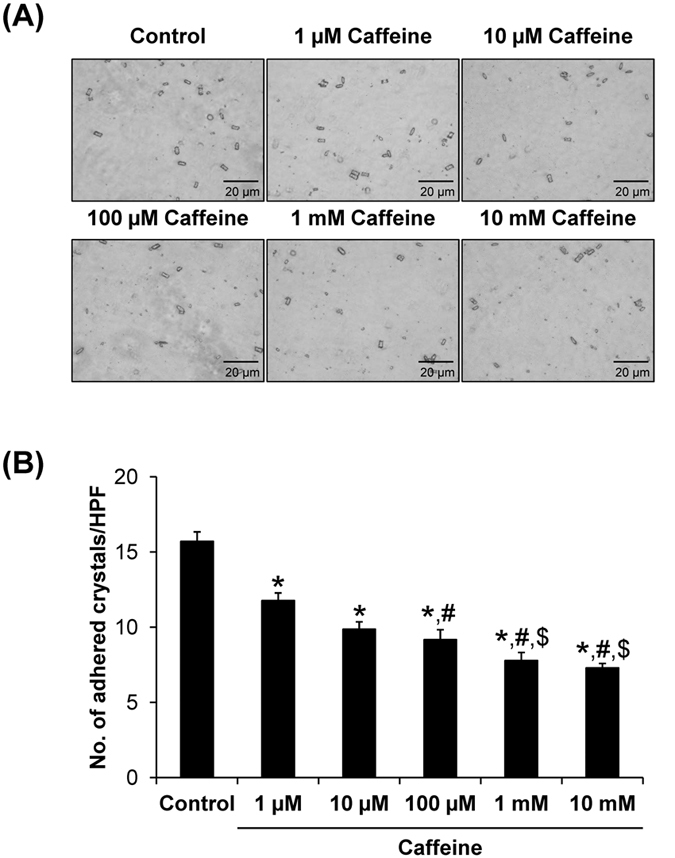
Figure 3. Cell-crystal adhesion assay.

(A) MDCK cells were pre-incubated with various doses of caffeine for 3 h prior to cell-crystal adhesion assay (the cells pre-incubated with no caffeine served as the control). Original magnification was 400× for all panels. (B) The adherent crystals remained on the cell surface after vigorous washes were counted from at least 15 HPFs. Each bar represents mean ± SEM from 3 independent experiments. *p < 0.05 vs. control; #p < 0.05 vs. 1 μM caffeine; and $p < 0.05 vs. 100 μM caffeine.
