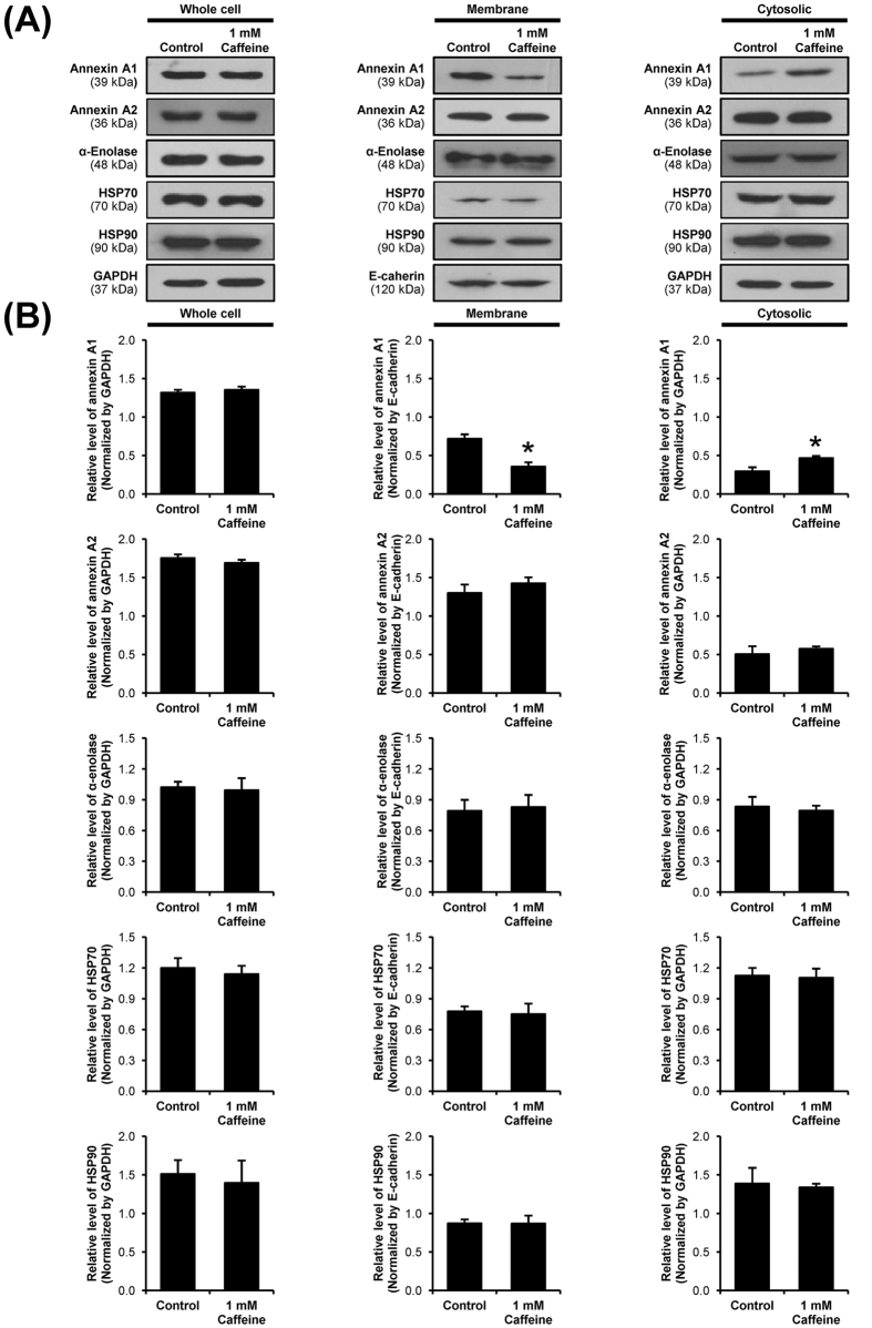
Figure 4. Western blot analysis of known COM crystal-binding proteins.
(A) Proteins derived from whole cell lysate, membrane fraction, and cytosolic fraction of the cells without or with 1 mM caffeine treatment were subjected to Western blotting of annexin A1, annexin A2, α-enolase, HSP70 and HSP90. GAPDH served as the loading control for whole cell lysate and cytosolic fraction, whereas E-cadherin served as the loading control for membrane fraction. Full-length blots of these cropped images are presented in Supplementary Figure S1. (B) Band intensity was quantitated using ImageQuant TL software (GE healthcare). Each bar represents mean ± SEM from 3 independent experiments. *p < 0.05 vs. control.

