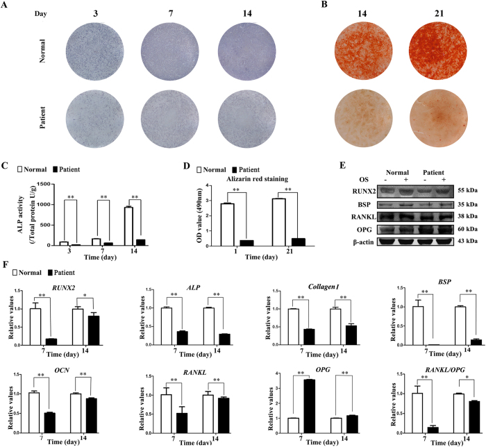
Figure 1. TDO-BMSCs exhibit weaker osteogenic potential than CON-BMSCs.
TDO- and CON-BMSCs at passage 3 were cultured under osteoinduction medium. (A) ALP staining assay on days 3, 7, and 14 after osteoinduction. (B) Alizarin red staining assay at 14 and 21 days post-osteoinduction. (C) ALP activity assay on days 3, 7, and 14 after osteoinduction. (D) Quantification of alizarin red staining on days 14 and 21 post-osteoinduction. (E) Western blots of RUNX2, BSP, RANKL, OPG, and β-actin protein 14 days after osteogenic stimulation. (F) Osteogenesis-related genes (RUNX2, ALP, COLLAGEN I, BSP, OCN, RANKL and OPG) mRNA expression after osteoinduction. GAPDH served as an internal control. Data were presented as the mean ± S.D. of 3 independent experiments. *p < 0.05; **p < 0.01.

