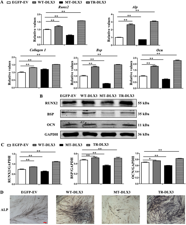
Figure 4. WT-DLX3 and TR-DLX3 increase while MT-DLX3 decreases cellular osteogenesis in MC3T3-E1 cells.
MC3T3-E1 cells were transfected with pEGFP-C1, pWT-DLX3, pMT-DLX3 and pTR-DLX3 respectively. After 24 h transfection, these cells were further cultured in osteoinduction medium for 72 h. Then, the cells were harvested and subjected to real-time PCR and western blot. (A) Osteogenesis-related genes (Runx2, Alp, Collagen I, Bsp, and Ocn) mRNA expression were determined by real-time PCR. GAPDH served as an internal control. (B) Protein expression of RUNX2, BSP, OCN, and GAPDH were detected by western blot. GAPDH served as loading control. (C) Quantification of protein expression related to GAPDH. (D) ALP staining assay after osteoinduction for 72 h was performed. Data were presented as the mean ± S.D. of 3 independent experiments. *p < 0.05; **p < 0.01.

