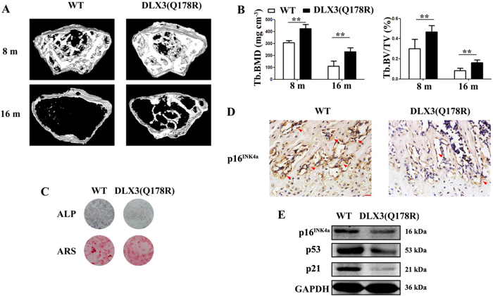
Figure 6. DLX3 (Q178R) attenuates skeletal aging and bone loss in vivo.
(A and B) Micro-CT analysis: 3D trabecular reconstructions (A) Trabecular BMD (Tb. BMD), Trabecular bone volume ratio (Tb. BV/TV) (B) of distal femoral metaphysis regions from 8- and 16-months-old WT and DLX3 (Q178R)-Tg mice. (C) ALP staining and Alizarin red staining (ARS) of femur bone BMSCs from 8-months-old DLX3 (Q178R)-Tg vs WT mice, after osteogenic induction. (D) Immunostaining with p16INK4a surrounding the trabecular bones from 8-months-old WT and DLX3 (Q178R)-Tg mice. Scale bars, 25 μm. (E) Western blot of p16INK4a, p53 and p21 in tibia and femur of 8 months mice.

