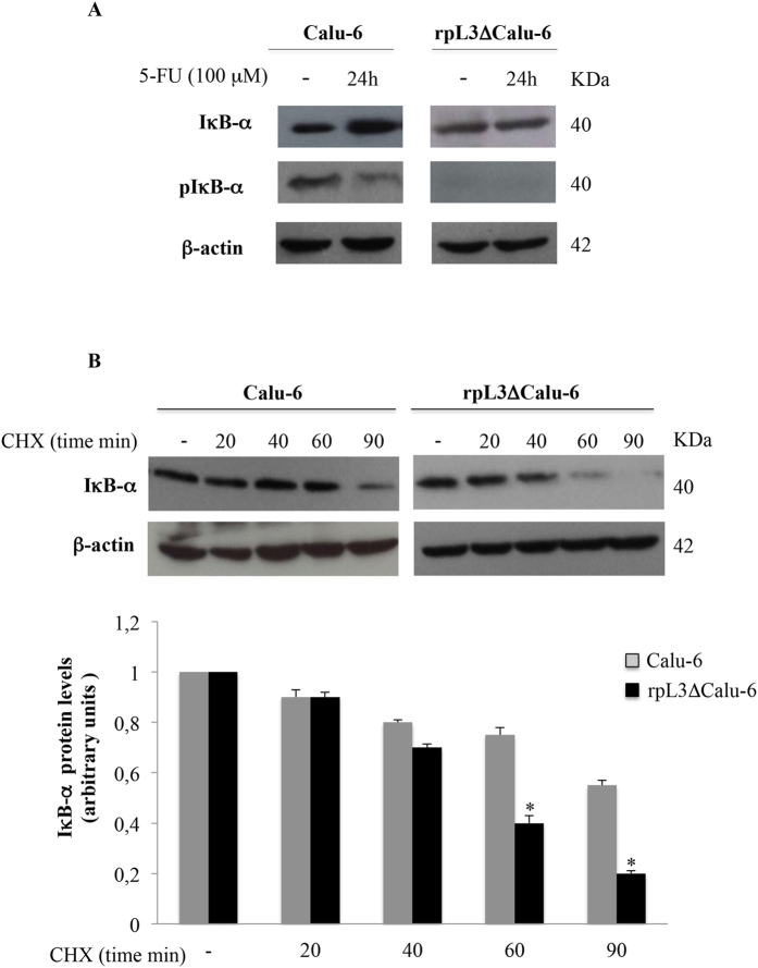
Figure 5. rpL3 prevents the degradation of IκB-α protein upon 5-FU treatment.
(A) Representative western blotting of IκB-α and pIκB-α expression. Calu-6 and rpL3ΔCalu-6 cells were treated or not with 100 μM 5-FU for 24 h. After the treatment, protein extracts from the samples were analyzed by western blotting with antibodies against IκB-α, pIκB-α and β-actin as control. (B) Calu-6 cells and ∆rpL3Calu-6 cells were treated with CHX for 20, 40, 60 and 90 min. Then, cell lysates were immunoblotted with anti- IκB-α and anti-β-actin antibodies. Quantification of signals is shown. *P < 0.05 vs. untreated ∆rpL3Calu-6 cells set at 1.

