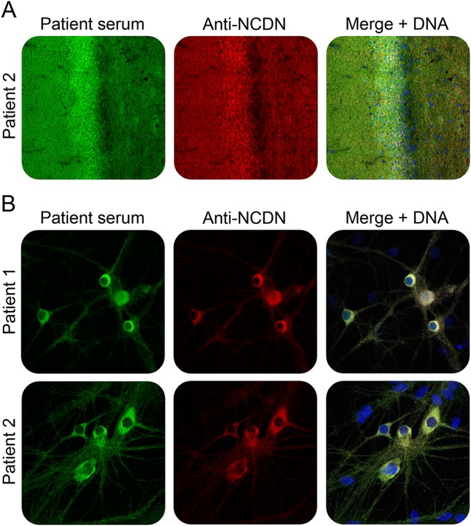
Figure 3. Double-staining of hippocampal tissue and cells with patient serum and rabbit antineurochondrin antibody.

Immunofluorescence staining of rat hippocampus tissue section (A) or formalin-fixed and TritonX-100 permeabilized rat hippocampal neurons (B) with patient sera (green) and antineurochondrin antibody (red). Nuclei were counterstained by incubation with TO-PRO-3 iodide (blue). (A, B) ×200 magnification.
