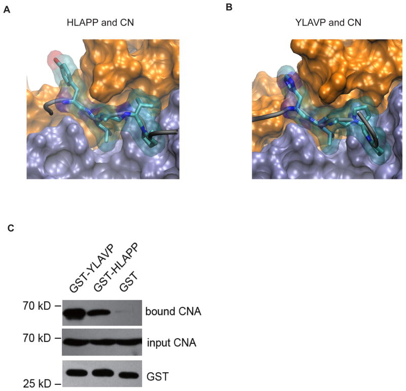
Figure 2. The binding between CN and the HLAPP motif of RCAN1 is weaker than that between CN and the YLAVP motif of NFAT.
In MD simulations the HLAPP (A) and YLAVP (B) motifs bind to CN in a similar manner. Both CNA (iceblue) and CNB (orange) are shown in surface presentation, while the LxxP motifs are drawn as stick and transparent surface representations. (C) Comparison of the binding between CN-YLAVP and CN-HLAPP in the GST pull-down experiments.

