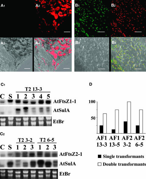
Figure 8.
35S:AtSulA-GFP Rescues 35S:AtFtsZ1-1 and 35S:AtFtsZ2-1 Induced Plastid Division Defects.
(A) and (B) Petals of single 35S:AtFtsZ transformants and of double 35S:AtFtsZ and 35S:AtSulA-GFP were observed using a confocal microscope. Bar = 20 μm.
(A) Petal of a 35S:AtFtsZ2-1 T3 progeny of the T2 6-5 single transformant. GFP fluorescence (A1), chlorophyll autofluorescence (A2), transmission (A3), and merge (A4).
(B) Petal of double transformants 35S:AtFtsZ2-1 and 35S:AtSulA-GFP from the same line. GFP fluorescence (B1), chlorophyll autofluorescence (B2), transmission (B3), and merge (B4).
(C) RNA gel blot. Transcript abundance of AtSulA and AtFtsZ1-1 in the progeny of the T2 35S:AtFtsZ1-1 line 13-3 retransformed with 35S:AtSulA-GFP (C1). C, Ws control; S, 35S:AtFtsZ1-1 single transformant. Transcript abundance of AtSulA and AtFtsZ2-1 in the progeny of the T2 lines 35S:AtFtsZ2-1 3-2 and 6-5 retransformed with AtSulA-GFP (C2). C, Ws control; S, 35S:AtSulA-GFP single transformant. AtSulA transcripts appear longer in transgenic plants because of the GFP fusion.
(D) Comparison between the proportion of WTL plants in double transformants and single transformants. Leaves of eight plants from each category were observed using an epifluorescence microscope.

