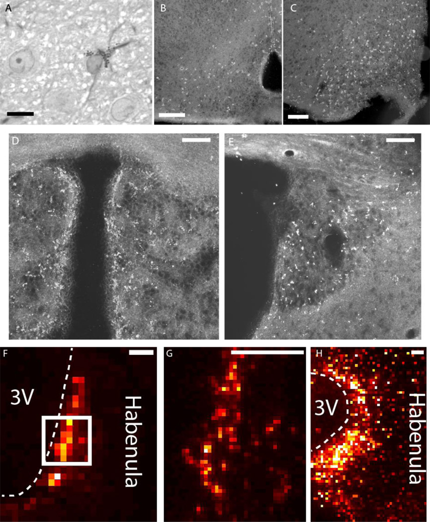
Figure 3.
A. High magnification view of a DAB-stained astrocyte, stained for GFAP using immunocytochemistry and counterstained with toluidine blue. An astrocyte possesses a cloud of DAB+ aggregates and GFAP-immunoreactive filaments surrounding an ovoid, pale-staining nucleus. Scale bar: 10 µm. B-E. Dark field micrographs for DAB-stained Gomori-positive astrocytes in frozen sections. DAB+ aggregates can be found in the arcuate nucleus and periventricular zone in the hypothalamus of both mice (B) and rats (C). The habenula in both mice (D) and rats (E) also exhibit similar distributions of DAB staining. DAB stain appears bright under Dark field microscopy. F-H. Cu XRF maps of mouse (F,G) and rat (H) habenulae demonstrating elevated Cu along the third ventricle. The box in F outlines the region imaged in G. Scale bars: 100 µm.

