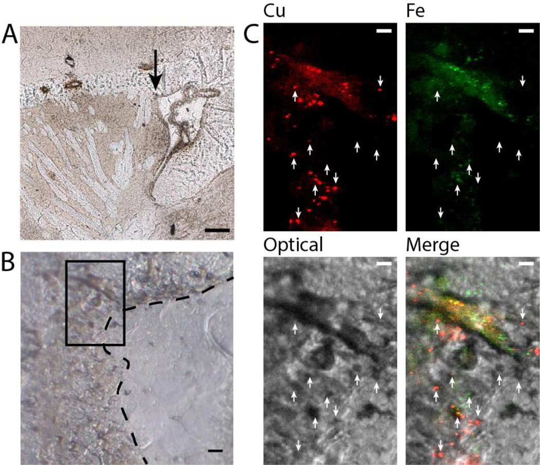
Figure 4.
A. Low-magnification micrograph of the DAB-stained section. The arrow points to the rostral corner of the ventricle, which was imaged. Scale bar: 100 µm. B. Untransformed optical micrograph of the rostral corner. The dashed line indicates the ventricle wall and the box outlines the area imaged with X-ray microscopy. Scale bar: 10 µm. C. Co-localization of Cu, Fe, and optical images showing that DAB+ aggregates often, but not always, co-localize with metal aggregates. Note that the optical image has been transformed as described in the methods section. Scale bar: 5 µm.

