Figure 1.
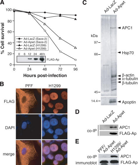
Apoptin is associated in vivo with APC1. (A) Saos-2 and H1299 cells were infected with adenovirus expressing Flag-Apoptin (Ad-Apwt) or LacZ (Ad-LacZ), and cell death was monitored for 96 h postinfection by trypan blue exclusion. (Inset) Immunoblot analysis using an α-Flag antibody to monitor Apoptin protein expression in Saos-2 cells. (B) Immunofluorescence of PFF and H1299 cells 24 h postinfection with Ad-Apwt. Cells were stained with an α-Flag antibody (left) or DAPI (middle); merged images (right). Magnification, 1000×. (C) Affinity purification of Apoptin-associated proteins from H1299 cells infected with Ad-Apwt. Proteins were separated by SDS-PAGE and visualized by silver staining. Microsequenced bands are indicated. (D) Apoptin was immunoprecipitated from Ad-Apwt-and Ad-LacZ-infected H1299 cells using an α-Flag antibody, and the immunoprecipitates were analyzed for APC1 by immunoblotting with a polyclonal α-APC1 antibody. (E) Apoptin was immunoprecipitated from Ad-Apwt-infected PFF or H1299 cells using an α-Flag antibody, and the immunoprecipitates were analyzed for APC1 by immunoblotting. Immunoblotting for Flag-Apoptin was performed on whole-cell extracts.

