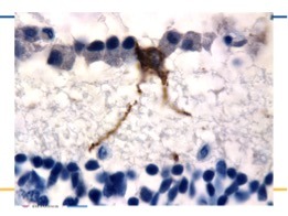
FIGURE 2.

MELANOPSIN RETINAL GANGLION CELL
A melanopsin retinal ganglion cell (brown) positioned along a line of normal retinal ganglion cells (blue) in a normal human retina. Note the extensions (dendrites) that are knobby, beaded and full of melanopsin which course down towards the inner nuclear layer.
