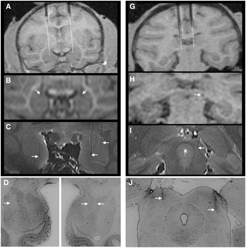
Fig. 2.
MRI and histological confirmation of infusion sites. (A) pre-infusion positioning of tungsten microelectrodes dorsal to the amygdala. Dotted line indicates the extension of the cannula calculated from the tip of the electrode. (B) Electrode tip positioned bilaterally in the amygdala. (C) Post-mortem ex vivo imaging showing close correspondence to ante-mortem scans. Infusion tracks are visible in both hemispheres and are identified by arrows. (D) Histological confirmation of infusion sites in the amygdala of a single subject. Minimal damage is evident. Tracks are indicated by arrows. (G) Pre-infusion positioning of tungsten microelectrodes dorsal to the superior colliculus. Dotted line indicates the extension of the cannula calculated from the tip of the electrode. (H) Electrode tip positioned in the superior colliculus. (I) Post-mortem ex vivo imaging showing a cannula track in the DLSC. (J) Histological confirmation of infusion sites in the DLSC of a single subject; tracks are evident bilaterally and indicated by arrows.

