Fig. 2.

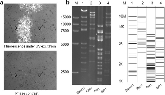
Plaques of mini-F recombinant PRV AH02LA strain and RFLP of BACPRV-G. a Images of mini-F recombinant PRV AH02LA and the parental AH02LA plaques under UV excitation (upper) and phase contrast (lower) are shown. Arrowhead shows a plaque of parental PRV AH02LA virus and arrow shows a plaque formed by mini-F recombinant PRV AH02LA. Each panel represents a view of 200 × 200 μm in size. b RFLP of BACPRV-G, DNA from PRV AH02LA BAC clone BACPRV-G was prepared by mini-prep and digested with BamH I, Kpn I, Pst I and Sph I (lanes 1–4). The digests were separated by 0.8% agarose gel electrophoresis for 16 h under 40 V (Left). Predicted RFLP patterns of BACPRV-G with BamH I, Kpn I, Pst I and Sph I digestion respectively. Predictions of these digestions were performed with the whole genome sequence of PRV ZJ01 strain (GenBank: KM061380.1) as reference. M: DL 15,000 DNA Marker (Takara)
