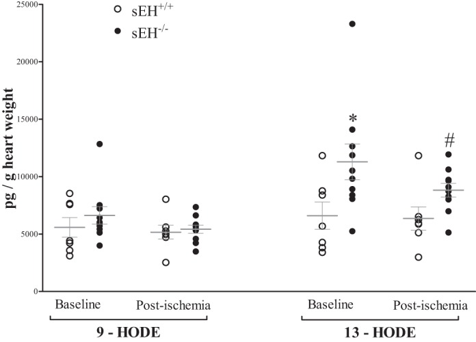
Fig. 6.

LC-MS/MS analysis of hydroxyoctadecadienoic acid (HODEs) in sEH+/+ and sEH−/− mouse heart perfusate samples at baseline (preischemia) and directly after 15-s ischemia (postischemia). Baseline and postischemia analysis 9- and 13-HODEs in sEH−/− vs. sEH+/+. 13-HODE, but not 9-HODE, was increased in sEH−/− vs. sEH+/+ mice at baseline and postischemia (P = 0.006). There was no change in either HODEs before and after ischemia in both groups (P > 0.05). *P ≤ 0.05 vs. baseline sEH+/+. #P ≤ 0.05 vs. postischemia sEH+/+. n = 7 sEH+/+, n = 10 sEH−/−.
