Fig. 7.
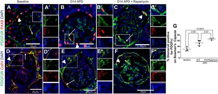
Marker for epithelial-to-mesenchymal transition PDGFβR is increased in PECs in APD mice given rapamycin. A–C: confocal microscopy showing immunofluorescent double staining for PDGFβR (green, arrowheads) and PAX8 (red, arrows) (magnification: ×400). PAX8 staining was used to demarcate PECs. Scale bars are provided. Nuclei were stained blue with DAPI. A: representative image of glomeruli at baseline showing PDGFβR in PECs. A′: a PEC positive for PDGFβR adjacent to a PAX8+ PEC is seen magnified and is shown as single-channel images below. B: the number of glomeruli positive for PDGFβR increases at D14 APD. The staining for PDGFβR on the same plane as PAX8 is apparent in the dashed box and is shown magnified in B′ and is shown as single-channel images to the right. C: representative image of glomeruli at D14 APD + rapamycin showing PDGFβR in PEC along Bowman's capsule. The staining for PDGFβR along PAX8+ PEC is apparent in the dashed box and is shown magnified in C′ and as single-channel images to the right. Collagen type IV staining was also used to demarcate Bowman's capsule, as there was no antibody cross reactivity. Scale bars are provided. Nuclei were stained blue with DAPI. We defined glomeruli with PDGFβR positive in PECs when there was PDGFβR signal on the urinary aspect of the collagen IV staining. D: representative image of glomeruli at baseline showing PDGFβR in PECs. D′: a PEC positive for PDGFβR but negative for collagen IV is seen magnified and is shown as single channel image below. E: the number of glomeruli positive for PDGFβR increases at D14 APD. The staining for PDGFβR on the urinary side of collagen IV is apparent in the dashed box and is shown magnified in E′ and is shown as single-channel images to the right. F: representative image of glomeruli at D14 APD + rapamycin showing PDGFβR in PEC along Bowman's capsule. The staining for PDGFβR on the urinary side of collagen IV is apparent in the dashed box and is shown magnified in F′ and as single-channel images to the right. G: graph showing percentage of glomeruli positive for PDGFβR along Bowman's capsule at baseline, D14 APD, and D14 APD + rapamycin was significantly higher at D14 APD + rapamycin compared with baseline and D14 APD.

