Fig. 1.
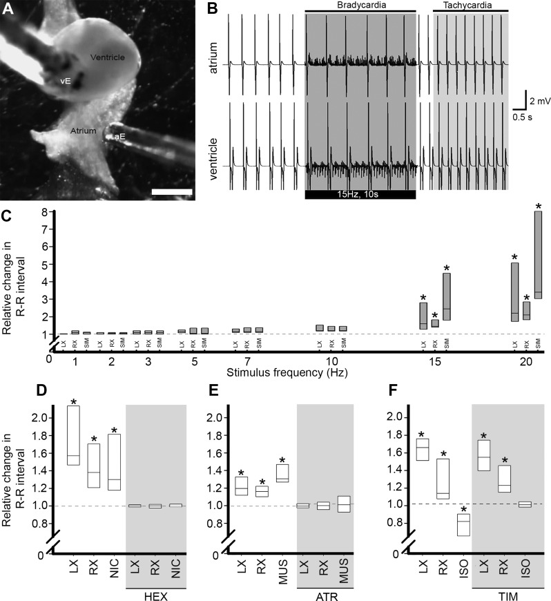
Chronotropic responses to vagosympathetic nerve stimulation and autonomic agents. A: in vitro view of the heart showing positions of bipolar electrodes recording atrial (aE) and ventricular (vE) surface electrocardiograms (ECG). Scale bar = 1 mm. B: typical ECG record from atrium (top) and ventricle (bottom) in the same heart before, during and after simultaneous stimulation of cardiac vagosympathetic nerve trunks (VSN; bar below trace). Elongation of R-R interval (bradycardia) during the stimulus period was followed by post-stimulus reduction of R-R interval (tachycardia) relative to prestimulus interval. C: proportional change in R-R interval in response to stimulation of the left (LX) or right (RX) VSN, or simultaneous stimulation of both nerves (SIM). *P < 0.05 vs. control by one-way ANOVA with Tukey's post hoc test; n = 8 for each. Dashed lines represent preintervention R-R interval. D–F: effects of cholinergic and adrenergic agents on R-R interval. D: R-R interval increased in response to LX and RX VSN stimulation and local application of nicotine (NIC). Effects of VSN stimulation and NIC application were eliminated during exposure to hexamethonium (HEX, grey shading). E: response to VSN stimulation and local application of muscarine (MUS) were eliminated by atropine (ATR, grey shading). F: isoproterenol (ISO) decreased R-R interval. Exposure to timolol (TIM, grey shading) blocked ISO response without affecting VSN-induced responses. *P < 0.05 vs. control by one-way ANOVA with Tukey's post hoc test; n = 8 for each of LX/RX/SIM and n = 6 for each of NIC/MUS/ISO.

