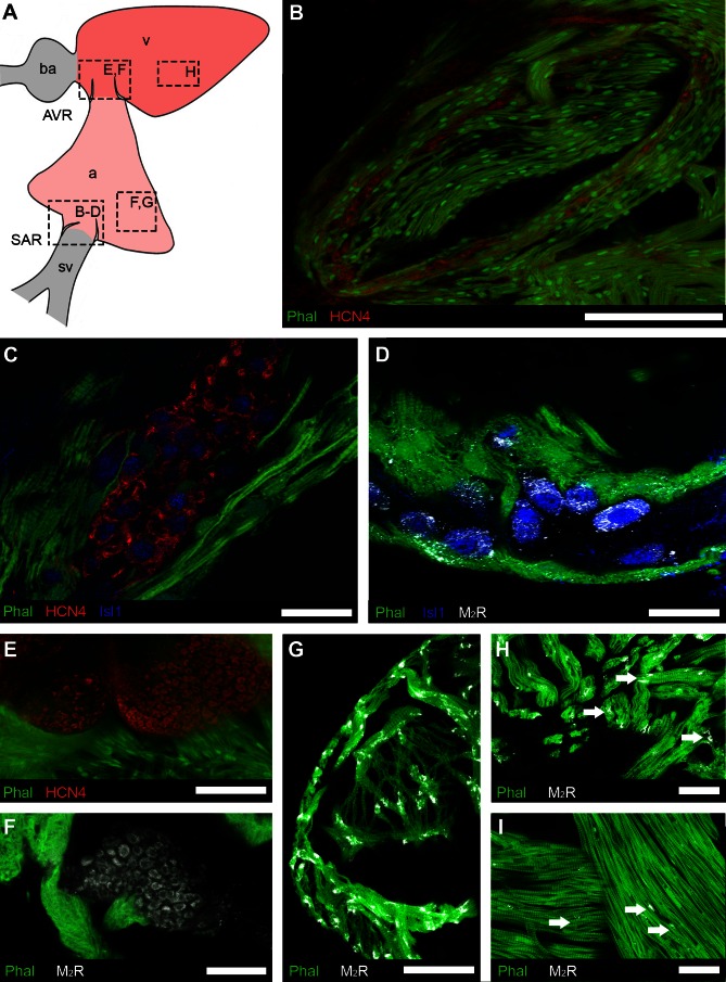
Fig. 4.
Regional distribution of putative pacemaker cells and muscarinic receptors detected by immunohistochemistry. A: schematic showing the cardiac regions: atrium, a; atrioventricular region, AVR; bulbus arteriosus, ba; sinoatrial region, SAR; sinus venosus, sv; ventricle, v. Boxes indicate the locations of images in B–H. B–D: organization of putative pacemakers and associated muscarinic receptors in SAR. B: low-magnification overview shows HCN4-immunoreactive (-IR) cells (red) embedded in musculature (Phal, green) surrounding the sinoatrial valves. C: islet-1 (Isl1, blue) co-localized with cells expressing HCN4. D: type 2 muscarinic receptors (M2R) appeared to be associated with the membrane of Isl1-IR cells. E: putative pacemaker cells expressing HCN4 were present in AVR, embedded in musculature (Phal) of atrioventricular valves. F: type 2 muscarinic receptors (M2R) associated with the membrane of cells of similar morphology to HCN4-IR cells in the AVR. G–I: M2R were more plentiful in the compact and trabecular myocardium (Phal) of the atrium (G and H; arrows) than in the ventricular trabecular myocardium (I; arrows). Scale bars = 200 μm (B), 40 μm (C), 20 μm (D), and 50 μm (E–I).

