Fig. 5.
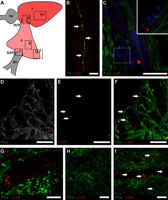
Regional distribution of β2-adrenergic receptors (β2-AR) associated with putative pacemaker cells and myocardium. A: schematic showing the cardiac regions; boxes indicate the locations of images in B–I. B: β2-AR (red) were detected adjacent to TH-positive innervation (green) in the atrial wall. C: in the sinoatrial region Isl1 (blue) was colocalized with cells expressing β2-AR. Inset shows a single confocal section from boxed region at a higher magnification. D–F: β2-AR-IR was detected in the compact and trabecular myocardium (Phal) of the atrium (E and F; arrows). G: β2-AR-IR associated with the membrane of cells of similar morphology to HCN4-IR cells in the AVR. H and I: β2-AR-IR was more plentiful in the compact myocardium (Phal) of the ventricle than in the atrium, especially proximate to coronary vasculature (I). Scale bars represent 10 μm (B), 40 μm (C), 20 μm (D–F), and 20 μm (G–I).

