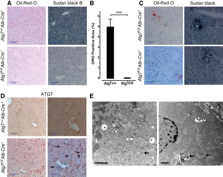
Fig. 5.
Liver fat stains, ATG7 stain, and TEM of Atg7F/FAlb-Cre+ mice. A: Oil-red-O (ORO) and Sudan black B (SBB) stain demonstrated a fasting-induced steatosis in Atg7+/+Alb-Cre+ mice (top), whereas lipids were virtually absent in Atg7F/FAlb-Cre+ mice (bottom). B: morphometric measurement of the ORO-positive area confirmed the virtual absence of ORO-positive lipids in the Atg7F/FAlb-Cre+ mice (Student's t-test, ***P < 0.001). C: representative photographs of ORO- and SBB-positive grouped cells in Atg7F/FAlb-Cre+ mice, which could be focally detected in the liver. These cells are mostly grouped around a larger droplet/vesicle and contain a mixture of larger and smaller lipid droplets seemingly arranged around the nuclei and plasma membranes. D: immunohistochemical staining with ATG7 confirms the knockout of Atg7 in Atg7F/FAlb-Cre+ mice, with absent cytoplasmic staining in the hepatocytes compared with Atg7+/+Alb-Cre+. The darker cells in Atg7F/FAlb-Cre+ mice compared with the surrounding cells appear to correspond to the cells with (condensed cytoplasm) proapoptotic features on the HE stain (see Fig. 3A). Larger magnifications demonstrate the positivity on the nonhepatocytes (arrows), whereas the ductular reaction is negative as well (as these cells also express albumin; arrowheads). Scale bars = 100 μm and 50 μm. E: TEM of the livers of Atg7F/FAlb-Cre+ mice. Some lipid-containing cells could be detected. These cells contained, not only lipid droplets, but also autophagic vacuoles, whereas lipid droplets and/or autophagosomes were not observed in the other hepatocytes. Arrows indicate autophagic vacuoles, asterisks indicate lipid droplets. Scale bars = 1 μm.

