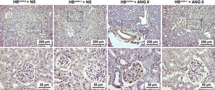
Fig. 6.
Representative images of VEGF-A staining in kidney sections from saline- or Ang II-infused mice. No obvious VEGF-A staining was detected in saline-infused kidneys in either group. In the kidneys from Ang II-infused mice, strong VEGF-A staining was seen in the glomeruli and vasculature in HBlox/lox control, which was decreased dramatically in the HBendo−/− kidneys.

