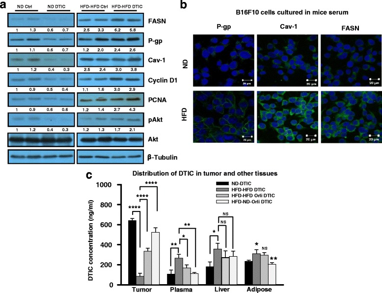
Fig. 2.

Molecular events associated with the impaired response to DTIC therapy in tumors of the experimental B16F10 isografted mice. a Western blot analysis of lysates from tumors of experimental HFD or ND mice subjected to SDS-PAGE and probed for levels of FASN, P-gp, Cav-1, pAkt, PCNA, and cyclin D1 in ND or HFD C57BL/6J mice treated with or without DTIC. b B16F10 or B16F1 cells were chronically grown in medium containing 5% serum collected from ND or HFD C57BL/6J mice for 15 days. Thereafter, these cells were subjected to immunofluorescence confocal staining of the indicated molecules (scale bar = 20 μm). c Quantitative determination of DTIC concentration in the tumor, plasma, liver, and adipose tissues excised from the indicated group of mice. Level of DTIC was determined by mass spectrometric analysis. The results are given as means ± standard deviation; *p < 0.05, **p < 0.01, ***p < 0.001, and ****p < 0.0001 denote significant differences between the groups; NS non-significant
