
Fig. 2.
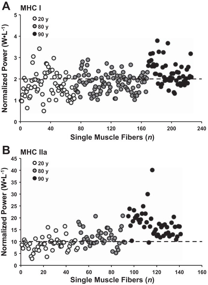
Normalized power (W/l) distribution of MHC I (A) and MHC IIa (B) single muscle fibers from 20-, 80-, and 90-yr-old individuals. Symbols represent individual muscle fibers from 20-yr-old healthy (n = 12, 6F/6M) (67); 80-yr-old healthy (n = 6M) (61); 90-yr-old healthy (n = 3F/2M) (current study). Dotted lines at 2 and 10 W/l represent average normalized power values in a large sample of older men and women (70–82 yr old) for MHC I and IIa fibers, respectively (61). All data were collected in the Human Performance Laboratory at Ball State University using identical procedures and instrumentation.