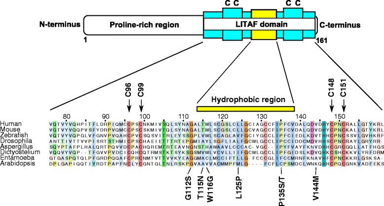
Fig. 1.

The domain organisation of LITAF. Schematic diagram to illustrate the domain organisation of the LITAF protein. The N-terminus is characterised by a proline-rich region which is followed by a LITAF domain at the C-terminus. The LITAF domain consists of two pairs of cysteine residues (indicated with arrows and coloured red) either side of a hydrophobic region. The amino acid sequences of selected LITAF domains taken from a variety of eukaryotes are shown to highlight the high degree of conservation of critical amino acid residues across species. Residues are numbered according to the human LITAF sequence and coloured according to the Clustalx scheme as implemented in the Jalview program to highlight conserved amino acid properties. Shading indicates the degree of conservation. The position of known CMT1C-associated pathogenic mutations are shown below the alignment.
