Fig. 6.

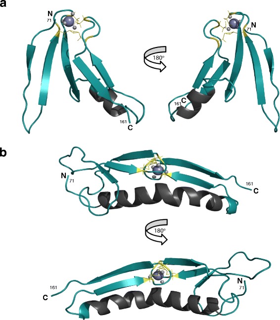
Rosetta structural models of LITAF. a Rosetta model of the C-terminal LITAF domain region of the LITAF Δ114–139 construct. The residue numbers correspond to the wild type human sequence. The zinc-coordinating cysteine residues are coloured yellow and the zinc atoms are illustrated by purple balls. b Rosetta model of the wild-type C-terminal LITAF domain. The zinc-coordinating cysteine residues are coloured yellow and the zinc atoms are illustrated by purple balls. The models calculated satisfy the known structural data: zinc coordination, NOE restraints and β-strand boundaries, with both models adopting a rubredoxin or zinc ribbon fold
