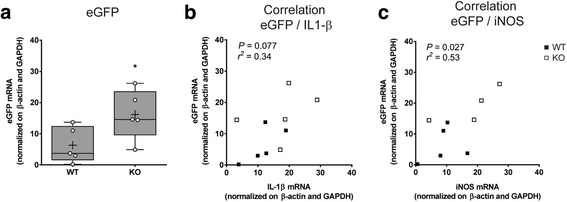
Fig. 3.

Tollip KO mice display a higher NF-κB-activation. Thirteen-month-old male mice underwent intra-nigral injection of rAAV9-NRE-eGFP (5 × 10⁸ viral genomes) followed by LPS (0.1 μg) injection 2 weeks later. Analyses were performed at 6 h post-injection of LPS (n = 5 mice per group). eGFP mRNA amounts in midbrain extracts were normalized by β-actin and GAPDH. a Results are represented as box and whiskers that show median, min., and max. values, 25th and 75th percentiles. Plots represent individual values and means are represented as a “+.” *P < 0.05 versus WT mice using a Mann–Whitney test. b–c Graph shows correlation between eGFP mRNA and IL-1β mRNA (b) and iNOS mRNA (c) in the injected side of Tollip KO (empty square) and WT (filled square) mice. The r squared was obtained using Pearson’s correlation
