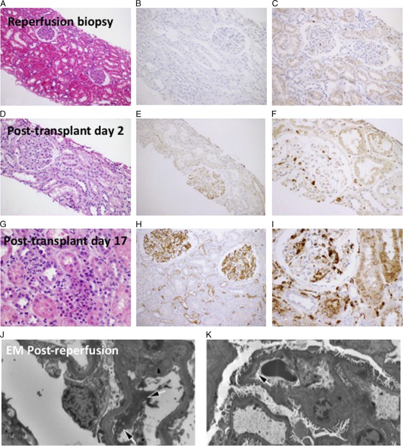
FIGURE 3.

Kidney transplant biopsies. Postreperfusion kidney transplant biopsy shows (A) mild tubular dilatation without significant inflammatory infiltrates, (B) nonspecific focal glomerular C4d staining, and (C) scattered CD68-positive mononuclear cells in the interstitium and in the glomeruli. On PTD 2, the renal allograft biopsy demonstrates (D) mild tubular injury, (E) diffuse linear C4d staining along glomerular and peritubular capillaries, and (F) scattered CD-68-positive mononuclear cells in the interstitium and in the glomeruli. Renal allograft biopsy on PTD 17 shows (G) tubular dilatation with prominent cytoplasmic vacuoles, mild inflammatory infiltrates and capillaritis, (H) stronger C4d staining in glomerular and peritubular capillaries, (I) numerous CD68 positive cells. J, K, Electron microscopic examination of the postreperfusion kidney transplant biopsy obtained approximately 30 minutes postreperfusion. There are already degenerative changes in the endothelial cell layer of capillary loops with a significant separation of the endothelial cells from the basement membrane (arrows).
