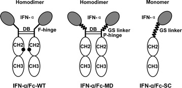
Fig. 1.

Schematic diagram for IFN-α/Fc fusion proteins. The dimer is composed of two molecules of IFN-α joined to dimeric Fc, and the monomer has a single molecule of IFN-α linked to monomeric Fc. Black circles N-glycosylation site in the wild-type IgG1 Fc region; DB disulfide bridges between dimeric Fc; F-hinge full hinge region of IgG1; P-hinge partial hinge with the amino acid sequence of HTCPPCP
