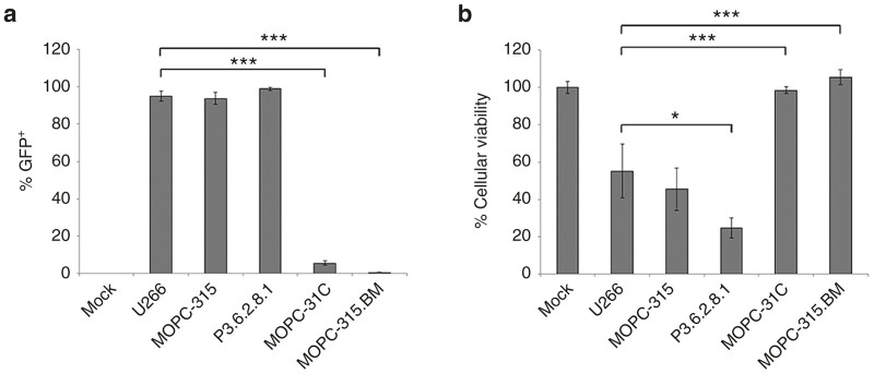
Figure 1.
MYXV infects murine MM cell lines. The indicated cell lines were mock infected or infected with vMYX-GFP at an MOI = 10. (a) Six hours postinfection, the number of GFP expressing cells was determined using flow cytometry. (b) Twenty-four hours postinfection, the effects of viral treatment on cellular viability was determined using MTT assay. Significance was determined using student’s t-test (*P < 0.05, ***P < 0.0001). GFP, green fluorescent protein; MM, multiple myeloma; MYXV, myxoma virus; MOI, multiplicity of infection.

