
Fig. 1.
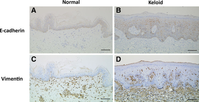
Immunohistochemical localization of E-cadherin and vimentin in representative samples of normal skin (A and C) and keloid tissue (B and D). The keloid keratinocytes exhibited greater vimentin expression (brown stain) than the normal keratinocytes. Bar, 100 μm.