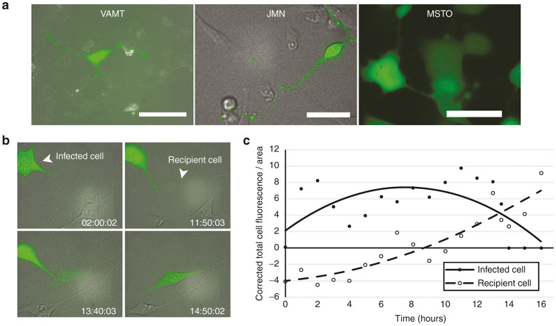
Figure 1.
TNTs mediate transfer of eGFP from a NV1066-infected mesothelioma cell to a noninfected mesothelioma cell. (a) NV1066-infected VAMT, JMN, and MSTO mesothelioma cells form TNTs following viral infection. Scale bars = 20 μm. (b) Time-lapse microscopy of a JMN mesothelioma cell infected with eGFP-expressing NV1066 forming a TNT that mediates eGFP transfer to a noninfected cell. This transfer took place over ~10–12 hours. (c) Quantification of eGFP expression in the infected (donor) and recipient cells from panel b as over time, reported using corrected total cell fluorescence per area (CTCF/Area). eGFP, enhanced green fluorescence protein; TNT, tunneling nanotube.

